
A research team in Japan, led by Nagoya University’s Akira Yokoi, has developed an innovative technique using cellulose nanofiber (CNF) sheets derived from wood cellulose to capture extracellular vesicles (EVs) from fluid samples and even organs during surgery. EVs are small structures from cancerous cells that play a crucial role in cell-to-cell communication. Extracting and analyzing EVs using this new technology has the potential to revolutionize early cancer diagnosis and open the door to personalized medicine. The researchers published their findings in Nature Communications.
Cancer is notorious for its poor prognosis and in many cases goes undetected until its advanced stages, leaving patients with limited treatment options. Detecting the cancer early using EVs and analyzing them provides vital information on disease status and its progression. This should assist physicians in monitoring and adjusting personalized cancer treatment plans. However, researchers have been limited in previous attempts to use EVs due to the lack of an effective isolation strategy.
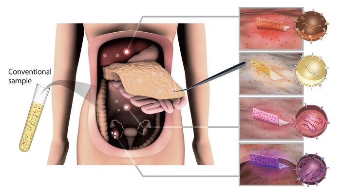
To capture EVs, Yokoi and his colleagues used CNF sheets made from wood cellulose to extract them from fluid samples from ovarian cancer mice models. As the material has a porous nanostructure, the sheet absorbs the fluid containing the EVs into its pores and closes them upon drying. They found that the sheets captured and preserved EVs from as little as ten microliters of body fluids. In contrast, the current standard methods, such as ultra-centrifugation, are more time-consuming and require much larger samples.
“We have developed the unique cellulose nanofiber by applying paper-making and solvent displacement technology,” Yokoi said. “The cellulose nanofibers we use are a sustainable biomass material that comes mostly from wood cell walls. These sheets have attractive properties, such as being lightweight, high strength, and most importantly, easily biodegradable.”
Using the technique, the researchers successfully extracted and analyzed EVs, and the microRNAs (miRNAs) contained within them from the mice ovarian cancer models. As miRNAs differ between healthy and sick patients, they represent an ideal diagnostic marker for cancer. They also identified distinct sets of miRNAs in EVs collected from tumor surfaces, some of which decreased after tumor removal. Tracking the presence or absence of these miRNAs could be an easy way to analyze the effectiveness of treatment and then tailor treatment according to tumor heterogeneity. Heterogeneity is a common problem where even in a single tumor, cancer cells have different characteristics and properties.
The structure of the sheets is similar to that of medical gauze, so they can easily be attached and removed even when placed on organs during surgery. To test this, the group used recently removed human organs. Their successful test revealed an exciting discovery, as the EVs on the tumor surface showed unique miRNA profiles compared to the tumor tissue.
“Organ surfaces were a previously unanalyzed EV subpopulation, which can now be subjected to biological assessments,” Yokoi said. “CNF paper enables the obtaining of EVs from multiple sites in the body. Then, by checking the molecular profiles of these EVs, we can monitor disease progression and tailor the selection of the best drug, contributing to personalized medicine.”
Dr. Takahiro Ochiya, Board Member of the International Society for Extracellular Vesicles and President of the Japanese Society for Extracellular Vesicles is enthusiastic about the potential of the sheets: “Exosome analysis using CNF sheets is an extremely novel method and is expected to have a variety of applications, including medical uses. We expect this to be a major advance that will bring the knowledge of exosomes as medical research directly to patients.”
This research has broad implications, opening up the analysis of EVs during surgery, an unexplored area until now. Looking ahead, the research team is committed to advancing the medical applications of EV sheets for various diseases, improving diagnostic accuracy, and helping usher in the era of personalized medicine.
Journal
Nature Communications
Article Title
Spatial exosome analysis using cellulose nanofiber sheets reveals the location heterogeneity of extracellular vesicles
Article Publication Date
8-Nov-2023