
Radiotherapy, which uses high-energy radiation to kill cancer cells, typically follows a dosing and treatment plan based on clinical research to minimize the impact of the radiation on healthy tissues. However, since the effects of radiotherapy can only be evaluated after the treatment has been completed, it is not possible to predict the immediate effects of the treatment on a patient or the potential side effects that might develop at a later stage because of the treatment. Since the effects of cancer treatment are highly variable, even conventional doses administered to patients can potentially cause long-term side effects in some people.
“Cancer therapies produce extensive changes in the physiological and morphological properties of tissues, which are also dependent on the individual,” says Associate Professor Teemu Myllylä, at the University of Oulu, Finland, who is part of a team built together with Chief Physicist, Dr. Juha Nikkinen, in the clinical medical physics division of radiotherapy at Oulu University Hospital, working to change the way cancer is treated by centering the treatment around the patient.
In a study published in Journal of Biomedical Optics (JBO), the researchers report developing a method to measure an individual’s biological response to therapy enabling them to evaluate in real-time the impact radiotherapy has on a patient.
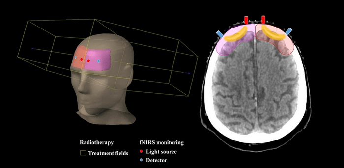
Their method involves the use of a functional near-infrared spectroscopy (fNIRS) device that uses infrared light to measure changes in the concentration of hemoglobin in the brain—an indicator of tissue oxygenation level. This noninvasive technique is commonly used to study brain activity in response to different stimuli and cognitive tasks. However, in this case, the researchers employed it to measure changes in the concentration of hemoglobin in the brain as the patient underwent whole-brain radiotherapy treatment where a linear accelerator was used to deliver targeted doses of radiation to the patient’s brain.
“A fiber optics setup using multiwavelength fNIRS was built and combined with a medical linear accelerator to measure cerebral tissue oxygenation changes during the brain radiotherapy treatment, where the radiation dose is limited to the brain areas to avoid irradiating the eyes,” says Myllylä.
Radiotherapy changes blood circulation in patient tissues and since hemoglobin levels are a useful indicator of changes in the blood volume, the use of fNIRS can provide insights into the effects of radiotherapy on blood circulation in the tissues. Using their technique, the researchers successfully measured tissue oxygenation levels in 10 patients and observed instantaneous changes in levels as the patients underwent multiple irradiations during their treatment.
“This is the first time human cerebral hemodynamics and cerebral tissue oxygenation changes have been measured during irradiation in clinical radiotherapy,” observes Myllylä. “The instantaneous measurement of tissue oxygenation levels during radiotherapy is especially helpful in cases of tumor hypoxia, which is when the oxygen levels in a tumor are low due to certain conditions. Such tumors are particularly resistant to radiotherapy. By instantly measuring tissue oxygenation levels, our proposed method can be used to assess the effectiveness of cancer treatment. This can eventually enable physicians tailor radiation doses to optimize treatment and improve outcomes for their patients,” he concludes.
The researchers plan to use their technique to measure tissue oxygenation levels to study how individual patients respond to different doses of radiation therapy. This can greatly help advance the field of personalized cancer treatment.
Read the Gold Open Access JBO article by T. Myllylä et al., “Cerebral tissue oxygenation response to brain irradiation measured during clinical radiotherapy,” J. Biomed. Opt. 28(1), 015002 (2023), doi 10.1117/1.JBO.28.1.015002
Journal
Journal of Biomedical Optics
DOI
10.1117/1.JBO.28.1.015002
Method of Research
Experimental study
Subject of Research
People
Article Title
Measuring changes in brain tissue oxygenation for personalized cancer radiotherapy
Article Publication Date
31-Jan-2023