Life Sciences
Modeling Disease in 3D
How do researchers, looking to create more realistic disease models—along with more targeted therapies—overcome the shortcomings of these models? Increasingly,…

The broad application of precision medicine has, until now, been largely hampered by the methods available to query the full complexity of humans and human disease. For instance, while immortalized cell lines have long been a workhorse of research in human biology, they are not able to reproduce the heterogeneity of diseases. In addition, the very process of immortalizing cells alters the fundamental biology of the tissue type they are derived from, and multiple propagations of cell lines introduce additional alterations. Mouse models, too, are limited, as they exhibit only a particular subtype of a disease and aren’t able to represent the diversity of disease phenotypes. Further, genetically engineered mouse models are widely considered poor predictors of whether a drug candidate will achieve the desired effects in humans.
So how do researchers, looking to create more realistic disease models—along with more targeted therapies—overcome the shortcomings of these models? Increasingly, it is via the use of human organoids. Research to create organoids—defined as miniature versions of organs that recreate the structure and biological complexity of an organ, along with its basic functions—dates back to the first half of the last century. But advances over the past dozen or so years in the methods for growing these three-dimensional models has resulted in a significant uptick in their use in precision medicine research.
Today, organoids grown from induced pluripotent stems cells, as well as from adult stem cells extracted directly from clinical biopsies, are being used for research into the basic biology of healthy human tissue, to understand disease progression, for drug screening in precision medicine research, and as potential therapies that can be transplanted to treat diabetes and other conditions. Lung organoids played a critical role during the pandemic as models to research SARS-CoV-2. Efforts are also underway to develop their use as a platform for new diagnostics that can help match patients to the most effective drugs.
As noted in a September 2021 paper in the Cell Press journal Med, “Using tissue derived from patients, these miniaturized models recapitulate various aspects of patient physiology and disease phenotypes, including genetic profiles and drug sensitivities. As such, patient-derived organoid (PDO) platforms provide an unprecedented opportunity for improving preclinical drug discovery, clinical trial validation, and, ultimately, patient care.”

Chief Scientific Officer
Tempus
According to Kate Sasser, PhD, chief scientific officer of the precision medicine company Tempus, the use of organoids for clinical research may also get a boost from regulatory bodies. “There is a lot of energy to move these forward,” Sasser says. “I think part of that is also galvanized now by the FDA Modernization Act, which is pushing the field towards reducing animal models and moving toward in vitro models. Organoids could potentially play a big role there.”
A better representation of cancer
Efforts to understand the genetic drivers of cancer and to use this information to help inform precision therapies have occupied the forefront of cancer research over the past 20 years. While there have been significant advances, so-called targeted therapies are still only effective in a subset of patients. Researchers understand that much of this is due to a variety of factors such as tumor heterogeneity, microsatellite instability, complex cellular interactions with the tumor, and the broader tumor biology. Genomic tests only capture one element of the disease, but with organoids—three-dimensional models that mimic the activity of the original tumor itself—researchers can better capture and analyze the diversity of cell types within the tumor, proteins, immune cells, and other markers of the disease. This more comprehensive view of cancer biology is today being applied to preclinical efforts to develop targeted therapies.
“When you do a clinical trial, we are always amazed at the heterogeneity of the responses” to a therapy, Sasser says. For example, designing therapies to rev up the immune system by targeting a protein called PD-1 that’s found on T cells “has been incredibly impactful in the clinic for certain patients, but not for others. Using these established biomarkers, we still are not able to uncover, or predict powerfully, which patients will respond, and which patients won’t.
“If you can get organoids to be reproducible for the human biology, then there are certain models now where you can include the immune system, you can include these other elements of the microenvironment like fibroblasts or stromal cells” that influence tumors, Sasser continues. Using techniques such as imaging, single-cell sequencing, proteomics, and flow cytometry on organoids, researchers can now better understand tumor biology at a cellular level to potentially develop new drug targets and unravel the mysteries of why some patients respond to drugs, while others don’t.
In particular, imaging applications for organoids that leverage the unique capabilities of artificial intelligence and machine learning are making an impact in the drug development arena by enabling screening of large numbers of drug candidates.
“You can imagine organoids at scale, where you have hundreds or thousands of organoids, and hundreds or thousands of drugs, and being able to apply imaging modalities to look at interactions between immune cells and tumor cells, or look at the different kinetics of tumor cell death, or look at really interesting endpoints that we that we wouldn’t necessarily capture in cell culture—like neighboring cells and how they’re interacting with each other,” Sasser says. “I think imaging is another important tool. Applying the machine learning modalities on top of that is uncovering a lot of new, exciting insights.”
Clinical applications
Ultimately, researchers and clinicians have an eye on bringing this new understanding of tumor biology into the clinic. At the University of Arizona College of Medicine, researchers including Yana Zavros, PhD, professor and associate head for research, are taking a translational approach to their work with organoids. A clinical trial designed by medical oncologist Rachna Shroff at the UA Cancer Center is leveraging patient-derived organoids to better understand mechanisms of resistance and to screen drugs for treatment response in metastatic pancreatic cancer.

professor
University of Arizona College of Medicine
“We’re learning about the tumor microenvironment through organoid generation of these patients on the clinical trial,” says Zavros, who leads the Biology Development and Research of Organoids (BioDRoid) service at the cancer center. “The advantage of using an organoid culture is now we can identify cells that are resistant to that therapy. The patient has been treated with the therapy, but we’re now able to isolate the cells that are resistant through an organoid culture, these are cells responsible for the high recurrence of disease in pancreatic cancer and metastasis. And we can use those organoids from these metastatic sites to screen for compounds that will target those cells.”
In addition to informing the development of drugs that are more effective at knocking out cancers, organoids also play a role in addressing the indiscriminate cytotoxicity of some cancer drugs and their resultant side effects. Curtis Thorne, assistant professor of cellular and molecular medicine at the UArizona College of Medicine–Tucson, has studied the complex interplay of cells lining the stomach and intestine, and how they maintain health, for the past six years. The Thorne lab focuses on how cells in the gut identify, measure, and respond to each other and to their environment; the signals that control the renewal and regeneration of tissues; and how some of the signals go haywire, leading to—and fueling—cancer. The lab’s research has leaned on the use of organoids for much of this work.
In a report published online by the University of Arizona Health Sciences, Thorne noted that organoids are a valuable tool to better understand the damage that can be done to the lining of the gut by cancer treatments.
“We are seeing that we can predict drug toxicity at a much earlier stage of the drug development. Generally, toxic compounds can be triggered early, and we can focus our efforts on those that are expected to have better toxicity profiles when tested in patients,” Thorne said in the report. “We think organoids are the best platform for demonstrating tumor behaviors in a dish in a form that we can screen drugs and identify better lead compounds that are more likely to work once they hit the clinic.”
Organoids also enable early pre-clinical work to be done at scale, Thorne added. “My lab views organoids as a drug-discovery platform. The way to think of them is as mini guts in tiny wells. We are able to put these miniature guts into 96- or 384-well plates. That allows us to do lots of experimental conditions to figure out how they grow and to discover new therapeutics that would prevent tumor organoids from growing.”
Scaling the technology
The increased use of organoids has spurred commercial development as IP derived from earlier research is spun out into companies that aim to further accelerate drug development, while also supporting clinical applications to help guide drug selection matched to a patient.
One such company is Xilis, which has pursued droplet technology to encapsulate patient-derived cells in order to miniaturize organoids into even smaller “organospheres.”

co-founder, CEO
Xilis
“By taking tissue from a biopsy, we can generate tens of thousands of little tumor avatars from a patient,” says company Co-founder and CEO Xiling Shen, PhD. “Now you have thousands of these patient tumor copies growing in these little droplets so you can do drug testing very quickly, to get back to the patient in ten to 12 days” with personalized information on the drug that will be most effective.
Research presented by Shen and colleagues in 2021 at ASCO showed that the organospheres created this way not only included the tumor cells, but also recapitulated the original, in vivo tumor environment. “You’re allowing not just tumor cells, but all the cells in the original environment—immune cells, the stromal cells,” he says. “Packed into that little environment, they create a mini ecosystem very quickly; you have the whole tumor microenvironment.”
There are other companies that are developing their own technologies to bring high-throughput, organoid-based screening platforms to research partners that can be used not only in preclinical work, but also in clinical trials. As Shen sees it, these are early days of collecting clinical data to directly prove the efficacy of high-throughput screening in predicting effective treatments.
“This allows pharma to explore treatment strategy before everything is locked down in Phase II, because you can explore different combinations and dosing strategy and do them together,” he said. “These are all very difficult to do in vivo, in a clinical trial.”
The future of organoids in the clinic
Before organoids can be developed and commercialized as a diagnostic tool, however, the technologies must be honed, automated, and made repeatable while researchers also gather data on sensitivity and specificity. Having the ability to conduct hundreds or thousands of experiments on the tumors of a single person in parallel will help speed the transition to their use in diagnostics. Shen believes the throughput speed for organoid-based systems will follow Moore’s law, providing a vast trove of clinically relevant data on a variety of cancer types.
Tempus is also preparing for a future where organoids can be used for diagnostic and prognostic insights. “The vision we can imagine is like a patient goes in, is diagnosed with cancer, and right from the beginning, organoid generation is part of the journey in their cancer care,” says Sasser. “Combined with other modalities like sequencing, we can understand what alterations are present from the very beginning, and what the best course of treatment could be.”
Chris Anderson, a Maine native, has been a B2B editor for more than 25 years. He was the founding editor for Security Systems News, and Drug Discovery News, and led the print launch and expanded coverage as editor in chief of Clinical OMICs, now named Inside Precision Medicine.
The post Modeling Disease in 3D appeared first on Inside Precision Medicine.
diagnostics
proteomics
therapeutics
stem cells
biomarkers
medical
pharma
artificial intelligence
machine learning
medicine
application
therapy
fda
research
clinical trials
clinical research


Wittiest stocks:: Avalo Therapeutics Inc (NASDAQ:AVTX 0.00%), Nokia Corp ADR (NYSE:NOK 0.90%)
There are two main reasons why moving averages are useful in forex trading: moving averages help traders define trend recognize changes in trend. Now well…

Spellbinding stocks: LumiraDx Limited (NASDAQ:LMDX 4.62%), Transocean Ltd (NYSE:RIG -2.67%)
There are two main reasons why moving averages are useful in forex trading: moving averages help traders define trend recognize changes in trend. Now well…

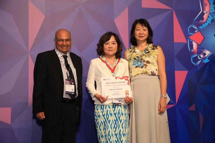
Asian Fund for Cancer Research announces Degron Therapeutics as the 2023 BRACE Award Venture Competition Winner
The Asian Fund for Cancer Research (AFCR) is pleased to announce that Degron Therapeutics was selected as the winner of the 2023 BRACE Award Venture Competition….