
Optical measurement techniques collecting light intensity in the far-field such as conventional and confocal microscopy or coherence scanning interferometry (CSI) enable fast and contactless inspection of several types of specimens. Nonetheless, optical measurement instruments suffer from diffraction effects leading to a fundamental lateral resolution limitation given by the minimum resolvable period length of Abbe-limit and the objective lens’s numerical aperture (NA).
In a new paper published in Light: Applied Manufacturing, a team of scientists led by Professor Peter Lehmann from the University of Kassel have developed a new model for modeling microscopic imaging under microsphere-enhanced interference microscopy.
Microspheres can be applied in microscopic imaging and measurement to overcome the resolution limit. Microspheres placed close to the surface are shown to enable a local improvement of the lateral resolution and a magnification enhancement. Microspheres can be combined with CSI to obtain electromagnetic phase information. Since the improvement of optical resolution is of great interest in many fields of application of microscopic imaging, microsphere-enhanced measurements are part of many recent experimental and theoretical publications.
Microspheres of high refractive index material can be combined with immersion objectives or embedded in elastomers. Microsphere-assisted measurements also apply to biological and medical objects such as viruses and sub-cellular structures or for identifying blood cells. As a result, microsphere-assisted measurements are used in many applications. Many theoretical studies are conducted to understand and analyze phenomena leading to resolution enhancement.
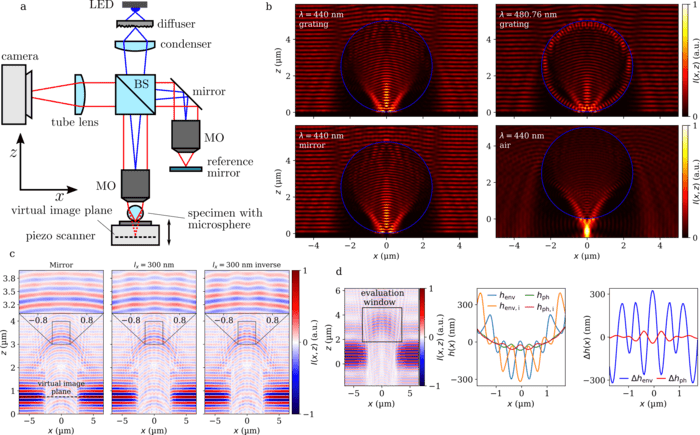
The research team has presented a simulation. It considers the full imaging process of a microsphere-enhanced interference microscope working in reflection mode equipped with objective lenses of high numerical aperture using a FEM calculation of the near-field scattering process. In contrast to previous theoretical models, they considered the full 3D conical Köhler illumination with incident waves and conical imaging of the scattered light field by the microsphere.
The model reliably reproduces measurement results, as demonstrated for several surface topographies measured with CSI. A first quantitative comparison with measurement results of microsphere-assisted interferometry is given. Using the model, the researchers have presented a method to qualify the resolution enhancement by a microsphere. They have demonstrated the relative improvement of the lateral resolution and have shown that the enhanced lateral magnification decreases with for high numerical apertures. In contrast, the field of view increases for larger NA values of the objective microscope lens.
Furthermore, the presented approach enables future researchers to analyze parameter influences and finding the most appropriate experimental setup depending on the shape, size, and material of the microelement as well as surrounding material to improve the resolution and profile fidelity of CSI. The model can be extended to conventional microscopy, confocal microscopy, and other optical profilers without significant effort. Therefore, the presented model can significantly contribute to a better understanding of microsphere-assisted measurement systems and improve their imaging capabilities through parameter studies.
Journal
Light: Advanced Manufacturing
DOI
10.37188/lam.2022.049