Medtech
Wearable Microscopes Image Spinal Cord Activity of Mice in Real Time
Scientists at the Salk have created wearable microscopes to enable deeper insight into the signaling patterns that occur within the spinal cords of mice….
While the spinal cord is known to play an essential role in relaying pain signals, technology has limited scientists’ understanding of how this process occurs on a cellular level. Despite progress in calcium imaging, which has enabled measurement of cellular activity, progress in developing other types of imaging has been an ongoing challenge.
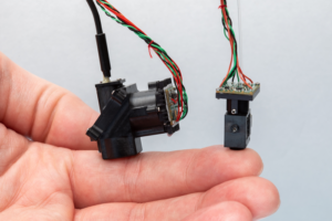
Now, scientists at the Salk have created wearable microscopes to enable deeper insight into the signaling patterns that occur within the spinal cords of mice. Using chronically implanted microprisms, they are able to image sensory and motor-evoked activity in regions, and at speeds, previously inaccessible.
This work may help researchers better understand the neural basis of sensations and movement in healthy and disease contexts—such as chronic pain, itch, amyotrophic lateral sclerosis, or multiple sclerosis.
This research is published in Nature Communications in the paper, “Multiplex translaminar imaging in the spinal cord of behaving mice.” The wearable microscope was first published earlier this month in Nature Biotechnology in the paper, “Trans-segmental imaging in the spinal cord of behaving mice.”

“These new wearable microscopes allow us to see nerve activity related to sensations and movement in regions and at speeds inaccessible by other high-resolution technology,” said Axel Nimmerjahn, PhD, associate professor, and director of the Waitt Advanced Biophotonics Center at the Salk. “Our wearable microscopes fundamentally change what is possible when studying the central nervous system.”
The wearable microscopes are approximately seven- and fourteen-millimeters wide (about the width of a little finger or the human spinal cord) and offer high-resolution, high-contrast, and multicolor imaging in real time. The new technology can be combined with a microprism implant, which is a small reflective glass element placed near the tissue regions of interest. This system, the authors noted, addresses multiple challenges of previous wearable microscopes, “including their limited working distance, resolution, contrast, and achromatic range.”
“The microprism increases the depth of imaging, so previously unreachable cells can be viewed for the first time. It also allows cells at various depths to be imaged simultaneously and with minimal tissue disturbance,” said Erin Carey, a research assistant in Nimmerjahn’s lab.

Pavel Shekhtmeyster, PhD, a former postdoctoral fellow in Nimmerjahn’s lab agrees: “We’ve overcome field-of-view and depth barriers in the context of spinal cord research. Our wearable microscopes are light enough to be carried by mice and allow measurements previously thought impossible.”
Nimmerjahn’s team applied the technology to gather new information about the central nervous system. In particular, they wanted to image astrocytes, star-shaped non-neuronal glial cells, in the spinal cord because previous work suggested a role for the cells in pain processing.
The team found that squeezing the tails of mice activated the astrocytes, sending coordinated signals across spinal cord segments. More specifically, they showed that dorsal horn astrocytes in behaving mice “show sensorimotor program-dependent and lamina-specific calcium excitation.” In addition, “tachykinin precursor 1 (Tac1)-expressing neurons exhibit translaminar activity to acute mechanical pain but not locomotion.”
Prior to the invention of the new microscopes, it was impossible to know what astrocyte activity looked like—or what any cellular activity looked like across those spinal cord regions of moving animals.
“Being able to visualize when and where pain signals occur and what cells participate in this process allows us to test and design therapeutic interventions,” said Daniela Duarte, a graduate student in Nimmerjahn’s lab. “These new microscopes could revolutionize the study of pain.”
Nimmerjahn’s team has begun investigating how neuronal and non-neuronal activity in the spinal cord is altered in different pain conditions and how various treatments control abnormal cell activity.
The post Wearable Microscopes Image Spinal Cord Activity of Mice in Real Time appeared first on GEN – Genetic Engineering and Biotechnology News.


ETF Talk: AI is ‘Big Generator’
Second nature comes alive Even if you close your eyes We exist through this strange device — Yes, “Big Generator” Artificial intelligence (AI) has…

Apple gets an appeals court win for its Apple Watch
Apple has at least a couple more weeks before it has to worry about another sales ban.
Federal court blocks ban on Apple Watches after Apple appeal
A federal appeals court has temporarily blocked a sweeping import ban on Apple’s latest smartwatches while the patent dispute winds its way through…